Le Neindre A, Philippart F, Luperto M, Wormser J, Morel-Sapene J, Aho SL, Mongodi S, Mojoli F, Bouhemad B. Int J Nurs Stud. 2021 May;117:103890. doi: 10.1016/j.ijnurstu.2021.103890. Epub 2021 Jan 29. Share this:

P H Mayo 1, R Copetti 2, D Feller-Kopman 3, G Mathis 4, E Maury 5 6 7, S Mongodi 8, F Mojoli 8, G Volpicelli 9, M Zanobetti 10 Affiliations 1Division of Pulmonary, Critical Care and Sleep Medicine, Northwell Health, Zucker School of Medicine at Hofstra/Northwell, New Hyde Park, Hempstead, NY, 11549, USA. mayosono@gmail.com. 2Department of Emergency Medicine, Latisana Hospital, 33053, Latisana, Italy. 3Division of Pulmonary, Critical Care, and Sleep Medicine, Johns Hopkins […]
Authors Mayo PH, Copetti R, Feller-Kopman D, Mathis G, Maury E, Mongodi S, Mojoli F, Volpicelli G, Zanobetti M. Intensive Care Med. 2019 Aug 15. doi: 10.1007/s00134-019-05725-8. [Epub ahead of print] Abstract This narrative review focuses on thoracic ultrasonography (lung and pleural) with the aim of outlining its utility for the critical care clinician. The […]
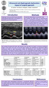
M Luperto, M Bouard, S Mongodi, F Mojoli, B Bouhemad Critical Care 2018, 22(Suppl 1):P211 Introduction: Diaphragm ultrasound (DUS) easily identifies diaphragmatic dysfunction (DD) in ICU patients [1], a potential cause of weaning failure (WF). Methods: Prospective observational monocenter study. We enrolled adults ICU patients at day 1 after surgery, extubated, with no neuromuscolar diseases. […]
F. Mojoli, S. Mongodi ICU Management & Practice. 2017;17(3):186-9. In the last years, ultrasound (US) became an essential tool in the hands of the intensivist and is now recommended both for procedural guidance and diagnostic purposes. Point-of-care ultrasound (POCUS) is an immediately available and repeatable, non-irradiating bedside tool integrating the clinical examination. While echocardiography […]
S. Mongodi, F. Mojoli, G. Via, G. Tavazzi, F. Fava, M. Pozzi, G. A. Iotti, B. Bouhemad
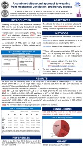
Introduction: Weaning failure (WF) from mechanical ventilation (MV) may be due to lung derecruitment, cardiac dysfunction and respiratory muscles weakness. Transthoracic echocardiography (TTE) [1], lung ultrasound (LUS) [2] and diaphragm ultrasound (DUS) [3] have shown their value in early identification of the failing patients separately. A combination of TTE, LUS and DUS could improve the identification of failing patients and of WF etiology [4]. We aimed to estimate the value of a combined ultrasound assessment (heart-lung-diaphragm) to early identify patients at high risk of WF from MV.
Methods: Prospective observational multicenter international study including all patients undergoing a 30’ spontaneous breathing trial (SBT) before extubation. Patients with neuromuscular diseases and MV <48 hours were excluded. TTE and LUS were performed before SBT and at its end; DUS at beginning and end of SBT. TTE included: MAPSE (mitral annulus plane systolic excursion), EF% (ejection fraction), E/A, E/Ea. We computed LUS score (0–36) [2]. DUS assessed diaphragm excursion. Extubation was considered as failed in case of reintubation, non-invasive ventilation or death within 48 hours. We used classical criteria for SBT failure. Extubation was decided by an independent operator.
Results: We enrolled 18 patients (age 77.7 ± 11.4 yrs, BMI 29.9 ± 7, SAPSII 54.4 ± 17.4, MV length 9.5 ± 7.3 days). 6 patients (33%) failed the SBT and were not extubated. 12 patients (67%) were extubated; 5 failed. Two populations were identified: WF (failed SBT or extubation) and weaning success (WS). LUS: SBT-LUS was higher than MVLUS in the whole population (17 ± 4 vs. 12 ± 3; p = 0.019); this was more remarkable in WF (20 ± 3 vs. 11 ± 3; p = 0.0004). SBT-LUS was higher in WF vs. WS (20 ± 3 vs. 13 ± 4; p = 0.03). SBT-LUS predicted SBT and weaning failure (respectively AUC 0,877 and AUC 0,883). TTE: E/A and E/Ea (both MW and SBT) did not predict WF, extubation failure nor SBT-failure. MV-MAPSE predicted WF (AUC 0,833); if < =10 mm it predicted WF with sensitivity 0.67 and specificity 1. SBTMAPSE predicted extubation failure (AUC 0.833). DUS: No correlation between diaphragm excursion and WF was identified.
Conclusions: LUS and MAPSE seem to be the most useful parameters to predict weaning failure.
References 1. Caille et al., Crit Care 2010 2. Soummer et al., Crit Care Med 2012 3. Kim et al., CCM 2011 4. Mongodi et al., Crit Care Med 2013
Grant The research project received the ESICM Clinical Research Award 2015.
Pag. 30 From Critical Care
Le Neindre A, Mongodi S, Philippart F, Bouhemad B J Crit Care. 2016 Feb;31(1):101-9. doi: 10.1016/j.jcrc.2015.10.014. Epub 2015 Oct 26 Abstract The use of diagnostic ultrasound by physiotherapists is not a new concept; it is frequently performed in musculoskeletal physiotherapy. Physiotherapists currently lack accurate, reliable, sensitive, and valid measurements for the assessment of […]

